Lichen Sclerosis Histology

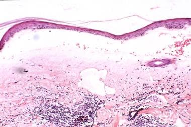
For the inflammatory phase of lichen sclerosus.
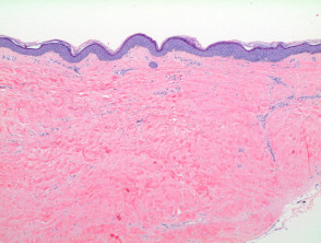
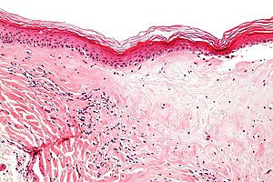
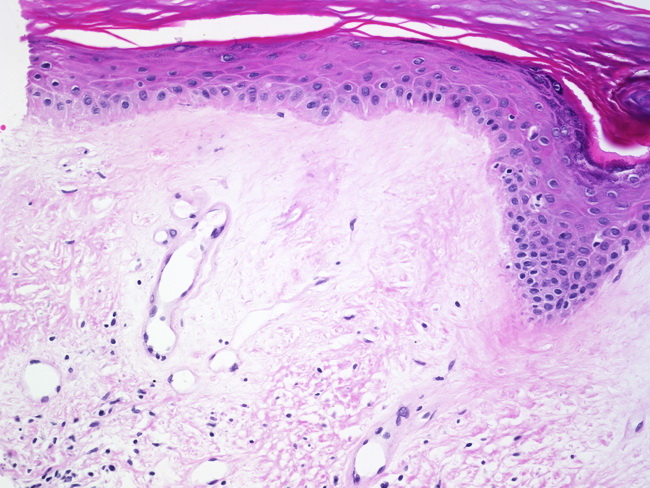
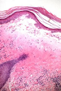
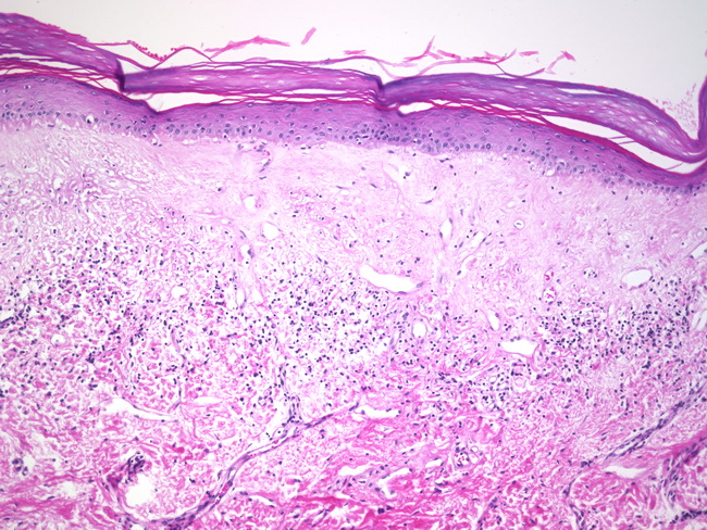
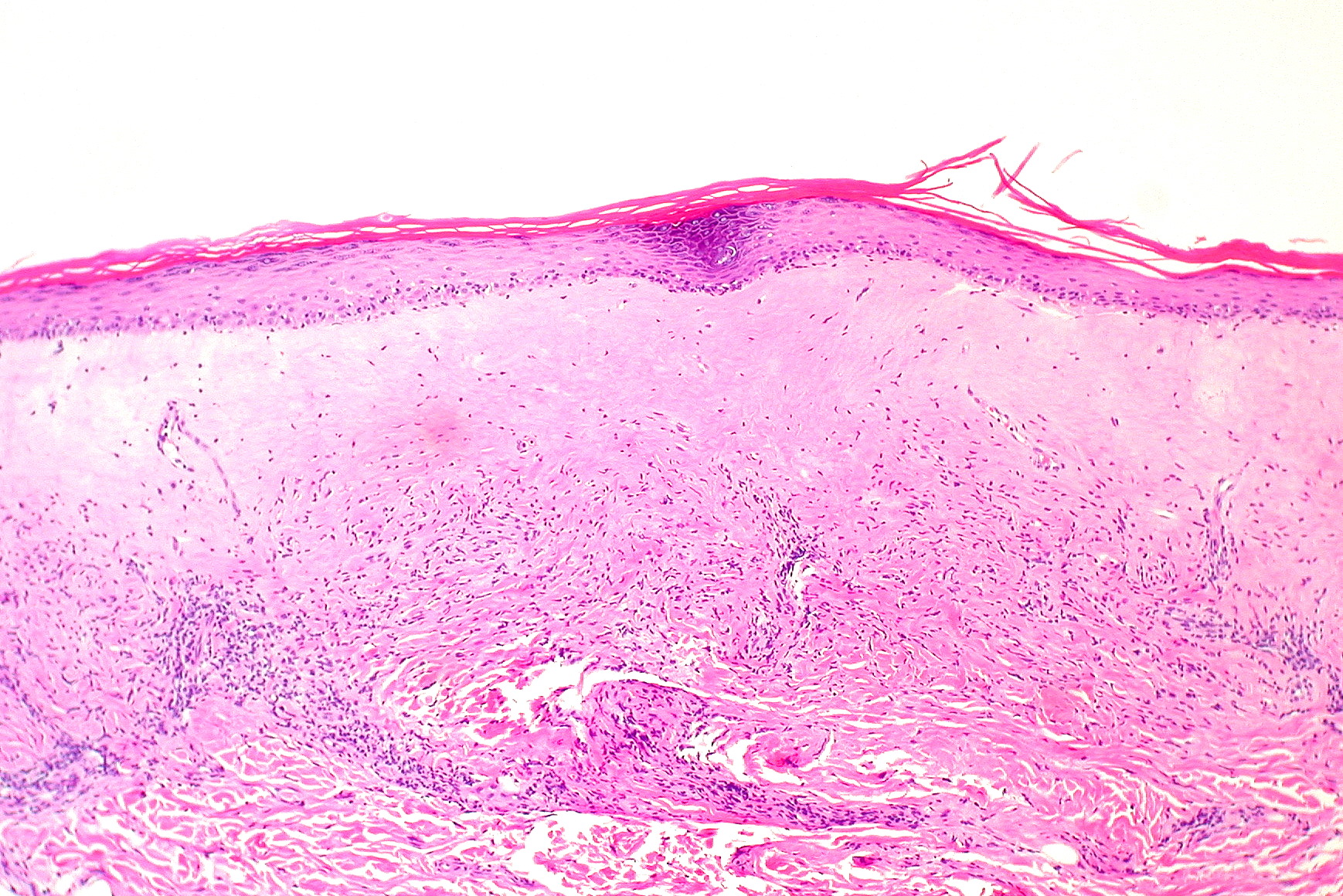
Lichen sclerosis histology. Lichen sclerosus ls is a relatively common dermatosis although the true prevalence of lichen sclerosus is unknown and likely underestimated. Scanning power of lichen sclerosus reveals a lichenoid inflammatory pattern in early stage lesions or a superficial sclerosing process in late stage lesions figure 1 and 2. Lichen sclerosus is a rare skin condition that usually shows up on your genital or anal areas. Lp has wedge shaped hypergranulosis lacks basilar.
May be confined to oral mucosa. Lichen sclerosus lie kun skluh row sus is an uncommon condition that creates patchy white skin that appears thinner than normal. Vulvar lichen sclerosus ls a lymphocyte mediated chronic skin disease begins with uncharacteristic symptoms and progresses undiagnosed to atrophy and destructive scarring. Anyone can get lichen sclerosus but postmenopausal women are at higher risk.
Self limiting lasting 1 2 years although longer for oral lesions. Differentiated vulvar intraepithelial neoplasia commonly co exists with lichen sclerosus. Some patients with longstanding advanced ls have an increased risk of vulvar carcinoma. This may be because patients are distributed among.
Early ls is treatable although not curable. The condition mostly affects adult women. Usually flexor arms and legs glans penis and mucous membranes. But it can also affect your upper arms torso and breasts.
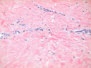
The epidermis shows hyperkeratosis significant thinning with loss of the normal rete ridge pattern and plugging of follicular infundibulae figure 3. Morphea profunda deep fibrosis. Lichen sclerosus ls is a chronic inflammatory skin disease of unknown cause commonly appearing as whitish patches on the genitals which can affect any body part of any person but has a strong preference for the genitals penis vulva and is also known as balanitis xerotica obliterans bxo when it affects the penis. Histology of lichen sclerosus.
